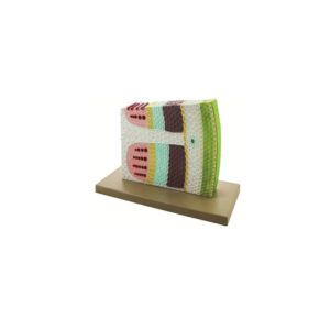
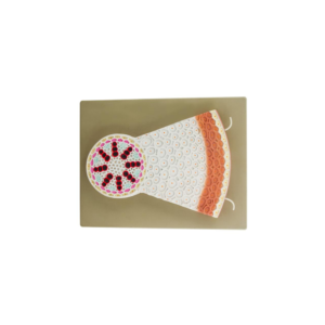
This model shows the transverse and longitudinal section of a dicotyledonous stem in which case the cambium ring has been formed but no secondary growth has yet taken place.
Useful model for study of epidermis, lentice , cork layer, cork cambium, cortical parenchyma ,starch sheath, medullary rays, phloem, sieve plate, sieve tube, phloem parenchyma and inter fasicular cambium ,xylem, pitted vessels, bordered pitted vessels, annular vessels, spiral vessels, pith etc.
Mounted on base, with key card.
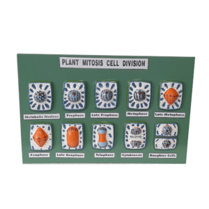
A set of 10 models showing all the stages of karyokinesis and cytokinesis from metabolic cell to the formation of two daughter nuclei.
All stages are mounted on a board with key card
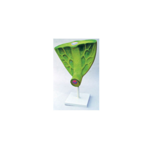
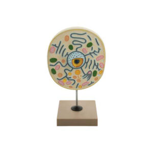
A highly enlarged model, as seen under electron microscope, a portion of wall is removed to show ectoplast, endoplast, tonoplast, vacuoles, nuclear structure, plastids, mitochondria etc.
mounted on base with key card.


Reviews
There are no reviews yet.