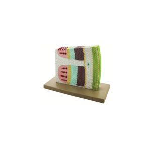
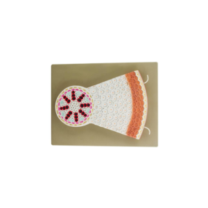
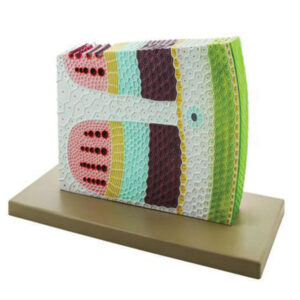
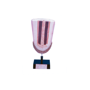
This model exhibits the various tissues and the scattered, closed and collateral vascular bundles in transverse and longitudinal sections in maize.
The large pitted vessels, spiral and annular vessels show the cell wall thickenings in L.S. Useful model for teaching anatomy of monocot stem.
Mounted on base with keycard.
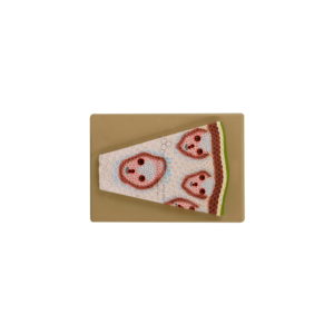
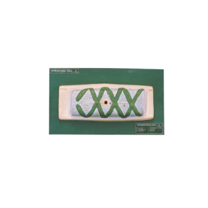
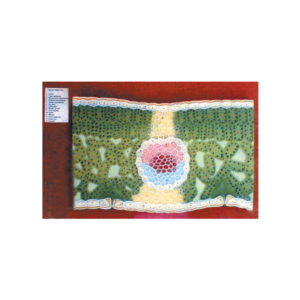
This model demonstrates the general arrangement of tissues. Stem is cut transversely, longitudinally, radially and tangentially at different planes to make the structural details very clear and the formation of various tissues easily understandable, mounted on stand with key card.


Reviews
There are no reviews yet.