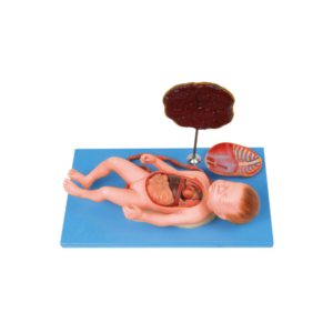
Demonstrate the whole process of delivery.
Demonstrate the fetus, umbilical cord and placenta of vacuum-assisted delivery, flexible fetal joint and multiple normal and abnormal positions of fetal delivery can be shown.
Can practice and master the comprehensive skills of normal labor, abnormal labor (dystocia), midwifery, and perineum protection.
Available Training of multiple pregnancies (twin pregnancy).
Component: A (Matrix for delivery Demonstration)* El (Fetus for Demonstration).
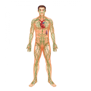
Lymphatic system model showing the different function of lymphatic system.
Relief model, approx. 2/3 natural size.
Model in one piece.
On a base plate.
Height: 84 cm., width: 54 cm., depth: 12 cm., weight: 10 kg
Suturing Simulators provide a realistic way for students to learn good surgical techniques.
The simulators provide students with a variety of repair experiences without the constraint of time and concern for safety, which are factors with a live patient.
The simulators can be used by the student in a learning lab with an instructor or by the student individually in the clinical setting just prior to a patient experience.
They are also useful as homework teaching aids that can be signed out at night and returned the next day.
In addition to being portable, the lifelike texture allows the learner to develop a “feel” for instrument handling, tension on suture, and the advantages of one method of tying knots over another.
Includes one of each simulator:
Midline Suture Simulator
Left Mediolateral Suture Simulator
Right Mediolateral Suture Simulator
Each simulator comes with an instruction booklet and is individually boxed.
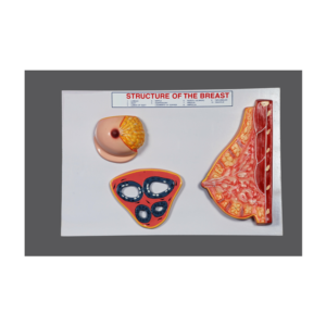
Demonstrate realistic self-examination with our natural casting of a female upper body with medium sized breasts.
It can easily be worn, in order to better train and practice breast self-examination.
Made of high-quality silicone.
Displays the skin in finest detail.
Very realistic to the touch and dermatologic ally tested
Breast examination is possible in both upright and lying positions.
Benign and malignant tumors in different stages of development hone self-examination skills:
2 benign tumors
4 malignant tumors
2 typical anomalies
Includes “Female Breast” chart
Supplied with talcum powder, harness, stand and aluminum carrying case.
For Continuous Ambulatory Peritoneal Dialysis.
Designed to introduce patients, students, and nurses to the essentials of CAPD procedures and care.
The realistic torso offers a realistic method to demonstrate and practice peritoneal dialysis.
Success with CAPD is dependent on the patient’s following dialysis procedures with extreme care.
This Life/form simulator gives the patient an opportunity to gain the confidence necessary for prolonged success with CAPD.
Complete with indwelling Tenckh off catheter in a hard carry case.
Dialysis materials not included.
The model is suitable for central venipuncture of internal carotid vein and subclavical vein and peripheric venipuncture (arm veins) of cephalic vein.
The cervical veins and arm veins can be filled with simulative blood.
Features:
The model is a simulative right half of adult torso with right arm.
There are apparent anatomical marks: clavicle suprasternal notch, stenocleidomastoid muscle, pectoralis major muscle, ribs and deltoid muscle.
Lifelike veins: Superior vena cava, internal jugular vein, subclavical vein, cephalic vein, basilic vein and median cubital vein etc.
With apparent anatomical marks, the model can be used to train puncture and catheterization of internal carotid vein, subclavical vein, cephalic vein and heart catheterization.
The skin and veins can be replaced, Students will feel an apparent “POP” when puncture needle has been thrust into the veins.
The heart floating swan-ganz intubation can be exercised
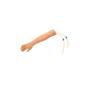
The I.V. Injection Arm P50/1 made of silicone, is unique in quality and design and allows realistic training to teach competence to medical staff.
It is also very suitable for group instruction because of its high quality, stain resistance and easy-to-clean soft material.
It is ideal for practicing the following:
Correct puncture of peripheral veins for blood sampling.
The following veins can be punctured: basilica vein, cephalic vein, median cubital vein, dorsal venous rete of hand
Intravenous injections
Positioning of a butterfly catheter
Delivered in deluxe storage carton with:
1 injection arm with already mounted tubing system
1 infusion bottle
1 stand
1 bottle of artificial blood concentrate(250ml)
1 plastic cup
1 disposable syringe and 2 injection cannulas (recommended cannula size: 20 and 21 gauge)
2 tubing systems as replacement parts
1 container of talcum powder


Reviews
There are no reviews yet.