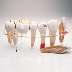
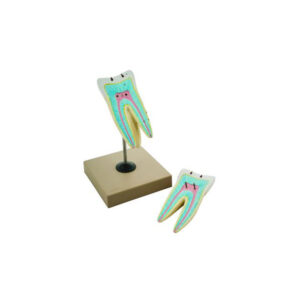
Human tooth,upper triple root but dissectible in 6 parts.
With removable pulp and three tooth inserts with different stages of advanced caries.
on stand with key card
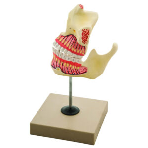
Upper triple root molar with caries, enlarged 15 times in 2 parts, longitudinal section through crown, 2 roots and pulp cavity, on stand with key card.
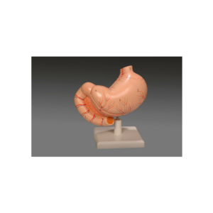
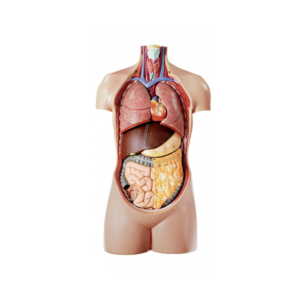
Full size,8 parts, shows all the major anatomical details.
Visceral organs are individually detachable to expose the body cavity.
Lungs, heart, liver, stomach and intestine are removable.
Heart, lung, and kidney are sectioned to show internal details, sexless, on base, with key card.
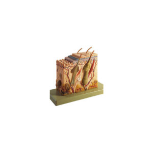
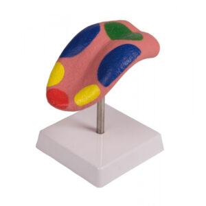
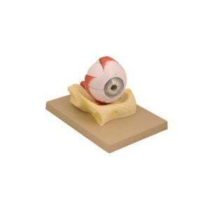
A 5 times enlarged model of the eye, sectioned horizontally, dissects into 7 parts.
The upper half of the sclerotic membrane, two choroid membranes, retina with vitreous humour, lens and lower half of the sclerotic membrane.
On base with key card.


Reviews
There are no reviews yet.