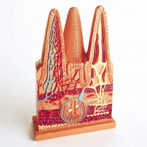
A highly improved and enlarged model showing fine details in three dimensional relief.
Cut in horizontal plane and separable in seven parts.
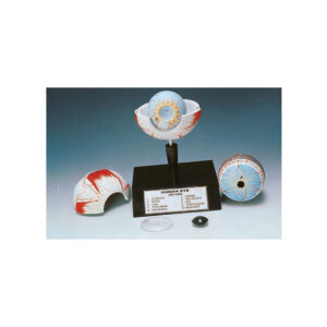
After removing the upper half of the sclera (outer shell).
The choroid with its verticose veins is exposed with the removal of second shell, details of the retina come into view and the position of the yellow and blind spots is quite visible. All important anatomical features such as muscle insertions, optic nerve, blood vessels, ciliary body, cornea, crystalline lens, and iris etc are clearly numbered and are identifiable. Mounted on base with key card.
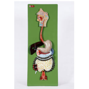
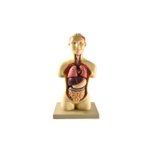
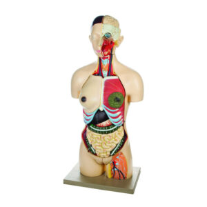
Youth model, approx. 600 mm. in height, dissectible into 6 parts.
The brain is fixed in skull and can be seen after removing the skull cap.
Lungs, liver, stomach and intestine are removable to show the internal structure.
On base with key card.
A 3 times enlarged model of the eye, with 9 parts, in bony orbit.
The upper half of eye ball is removable and dissects into 7 parts, showing all anatomical details.
Out of six muscles controlling the eye ball two are removable.
On base with key card.


Reviews
There are no reviews yet.