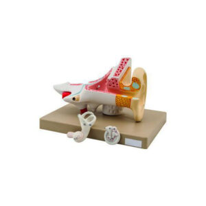
Two times enlarged, dissects into 3 parts, showing external, middle and internal portions.
Ossicles and labyrinth can be taken out.
On base with key card.
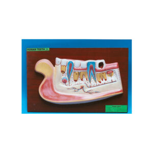
Greatly enlarged, internal wall is removed to show the incisors , molars, permanent canine and permanent premolars in full view, as also the arteries, veins and nerves serving them, on a large size board, with key card.
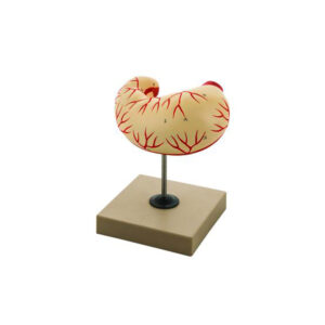
A 5 times enlarged model of the eye, sectioned horizontally, dissects into 7 parts.
The upper half of the sclerotic membrane, two choroid membranes, retina with vitreous humour, lens and lower half of the sclerotic membrane.
On base with key card.
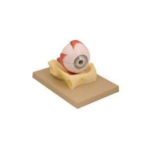
A 3 times enlarged model of the eye, with 9 parts, in bony orbit.
The upper half of eye ball is removable and dissects into 7 parts, showing all anatomical details.
Out of six muscles controlling the eye ball two are removable.
On base with key card.
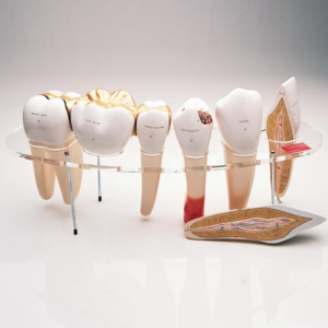
Upper triple root molar with caries, enlarged 15 times in 2 parts, longitudinal section through crown, 2 roots and pulp cavity, on stand with key card.


Reviews
There are no reviews yet.