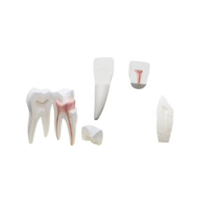
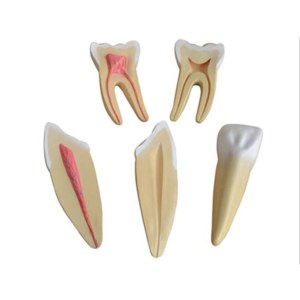
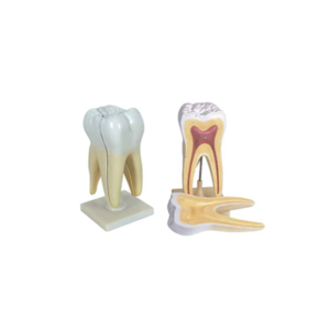
This model shows the morphological differences of the incisor, canine and molar teeth.
Dissection of the canine and molar teeth demonstrate the structure of enamel, dental pulp cavity.
Enlarged 12 times, 3 pcs set.
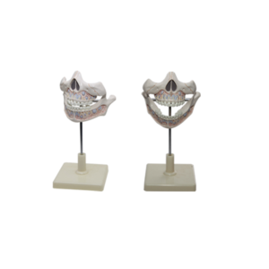
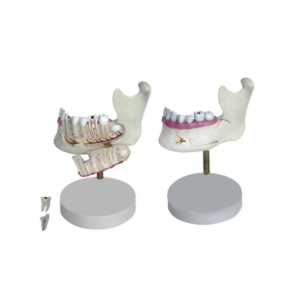
The model represents half of the left lower jaw of a young person,
One section of bone is removable to expose the tooth roots,
spongiosa,vessels and nerves.
Canine and first molar are removable and longitudinally sectioned.
On stand.
Size(cm): 14x17x15
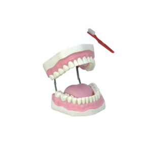
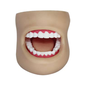
Model is making from the life size dental.
Anatomical structure including:
cheek, palatal, teeth(crown), gums, upper and lower dental arch, tongue.
It demonstrates teeth form and oral cavity cleaning and protection.
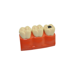
This two-sided teeth pathologies model by GPI Anatomicals shows tooth,gum and jaw structure and common pathologies including cavity decay,cracked tooth with cavity,perodotitis,plaque/tartar and more.
Excellent visual support for any dental hygiene curricul


Reviews
There are no reviews yet.