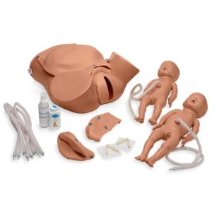
Demonstrate the whole process of delivery.
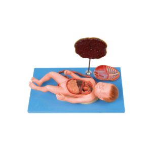
Demonstrate the fetus, umbilical cord and placenta of vacuum-assisted delivery, flexible fetal joint and multiple normal and abnormal positions of fetal delivery can be shown.
Can practice and master the comprehensive skills of normal labor, abnormal labor (dystocia), midwifery, and perineum protection.
Available Training of multiple pregnancies (twin pregnancy).
Component: A (Matrix for delivery Demonstration)* El (Fetus for Demonstration).
The model basing on the mechanism of normal labor shows the cardinal movements of engagement.
Descent-flection-internal rotation of the head-extension-retitution-external rotation of the head-internal rotation of the shoulders at accouchement.
Suitable for teaching the mechanism of normal labor in medical colleges and nursing schools.
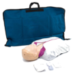
Demonstrate realistic self-examination with our natural casting of a female upper body with medium sized breasts.
It can easily be worn, in order to better train and practice breast self-examination.
Made of high-quality silicone.
Displays the skin in finest detail.
Very realistic to the touch and dermatologic ally tested
Breast examination is possible in both upright and lying positions.
Benign and malignant tumors in different stages of development hone self-examination skills:
2 benign tumors
4 malignant tumors
2 typical anomalies
Includes “Female Breast” chart
Supplied with talcum powder, harness, stand and aluminum carrying case.
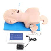
The model is suitable for central venipuncture of internal carotid vein and subclavical vein and peripheric venipuncture (arm veins) of cephalic vein.
The cervical veins and arm veins can be filled with simulative blood.
Features:
The model is a simulative right half of adult torso with right arm.
There are apparent anatomical marks: clavicle suprasternal notch, stenocleidomastoid muscle, pectoralis major muscle, ribs and deltoid muscle.
Lifelike veins: Superior vena cava, internal jugular vein, subclavical vein, cephalic vein, basilic vein and median cubital vein etc.
With apparent anatomical marks, the model can be used to train puncture and catheterization of internal carotid vein, subclavical vein, cephalic vein and heart catheterization.
The skin and veins can be replaced, Students will feel an apparent “POP” when puncture needle has been thrust into the veins.
The heart floating swan-ganz intubation can be exercised
With the female catheterization trainer, female bladder catheterization procedures can be realistically demonstrated, practiced and assessed.
The genital inserts are placed into the anatomically correct lower abdominal model and held in place with magnets.
The material is soft and flexible:
the foreskin is movable and the penis can be stretched.
The material gives a realistic feeling when inserting and removing the catheter into the bladder with realistic resistance and insertion depths.
Direct learning:
When the catheter has been correctly inserted, fluid runs out, just like with a real patient.
The catheter can also be checked through the transparent bladder.
The abdominal wall can be removed so that the process can be viewed better.
Includes suprapubic catheter:
If it is no longer possible to carry out catheterization through the urethra, a suprapubic catheter can be inserted.
On the PRO version, a suprapubic catheter has already been put in place, and the student can practice cleaning it and taking care of it with the trainer.
All the benefits of the female catheterization simulator at a glance:
Female catheterization with realistic resistance
Soft and movable labia (for practicing disinfection, for example)
Anatomically realistic pelvic structure
Liquid outflow if catheterization is successfully carried out
The transparent bladder can be checked and the abdominal wall can be removed.
Includes suprapubic catheter for practicing, cleaning and caring for it.
Stands securely on the table thanks to the non-slip feet
Magnetic connectors for quick set-up and dismantling
Easy to clean and maintain and can be completely taken apart
A sponge on the inside prevents a build-up of moisture
This model provides prostates with tumors in 4 different development periods.
The 1st development:
Beginning, a little big but the prostates still works well.
The 2nd development:
Some hard tubers can be served in the top right quadrant of prostate.
It's the beginning of the tumors.
The 3rd development:
The tumor is proliferating, and the previous tubers become bumps.
The 4th development:
The prostates is all replaced by tumors, the surface become more rough and harder than before.
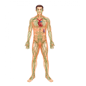
Lymphatic system model showing the different function of lymphatic system.
Relief model, approx. 2/3 natural size.
Model in one piece.
On a base plate.
Height: 84 cm., width: 54 cm., depth: 12 cm., weight: 10 kg

















Reviews
There are no reviews yet.